Summary
This article explains the Modified Beer-Lambert Law (MBLL), which adjusts for light scattering in tissue to calculate chromophore concentrations in fNIRS. It introduces the Differential Pathlength Factor (DPF) for total optical pathlength and the Partial Pathlength Factor (PPF) for brain-specific pathlength, highlighting their roles in accurate brain measurements.
Modified Beer-Lambert Law
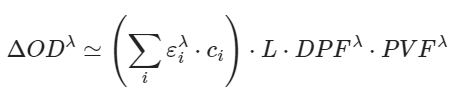
The modified Beer-Lambert law (MBLL) helps account for how light scatters as it moves through tissue, which increases the effective distance light travels. This effective distance is called the optical pathlength. The MBLL equation is:
In this equation:
-
ΔOD is the change in optical density (absorption) measured between a source and detector in an NIRS system.
-
εiλ is the extinction coefficient for a specific wavelength (λ) and chromophore.
-
ci is the concentration of that chromophore.
For fNIRS, the two main chromophores are HbO and HbR. While the original Beer-Lambert law assumed a straight optical path, the MBLL adjusts for scattering, which makes photons travel a longer, diffuse path in tissue. This adjustment is achieved using a unitless scalar called the differential pathlength factor (DPF).
How the DPF Works
The DPF adjusts the measured distance between the light source and detector (L) to account for scattering. For example, if the source and detector are 3 cm apart on the scalp (L), photons might travel an effective pathlength of ~18 cm because of scattering. In this case, the DPF is 6 (18 cm = 3 cm × 6).
However, much of this path (18 cm) is through layers like skin, skull, and cerebrospinal fluid (CSF), which are not the primary focus of fNIRS. To account for how much of the path is actually in the brain, we apply the partial volume factor (PVF).
Introducing the PPF
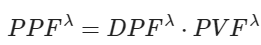
The partial pathlength factor (PPF) accounts for the effective pathlength in the brain specifically. The PPF is calculated by combining the source-detector distance (L), the DPF, and the PVF:
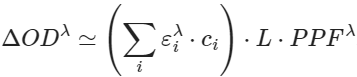
When applied to the MBLL, the brain-specific version becomes:
Key Difference: DPF vs. PPF
- The DPF adjusts for the total optical pathlength through all tissues (skin, skull, brain, etc.).
- The PPF adjusts specifically for the pathlength in the brain tissue.
This makes the PPF particularly important when studying factors like age, sex, or head size, as it directly relates to the signal changes measured in the brain. The optical density changes (ΔOD) measured by fNIRS are proportional to the PPF, making it a critical factor for accurate brain-specific measurements.
References
Cope, M., Delpy, D. T., Reynolds, E. O. R., Wray, S., Wyatt, J., & Richardson, C. E. (1988). Methods of quantitating cerebral near infrared spectroscopy data. Advances in Experimental Medicine and Biology, 222, 183–189. Springer. https://doi.org/10.1007/978-1-4615-9510-6_21.
Whiteman, A. C., Santosa, H., Chen, D. F., Perlman, S. B., & Huppert, T. J. (2018). Investigation of the sensitivity of functional near-infrared spectroscopy brain imaging to anatomical variations in 5- to 11-year-old children. Neurophotonics, 5(1), Article 011009. https://doi.org/10.1117/1.NPh.5.1.011009.
Webinar: Introduction to NIRS Toolbox - Installation & Getting Started offered by NIRx Medical Technologies on April 23th, 2020. Reference to the explanation given by Dr. Ted Huppert at this question starting from minute 41:30.